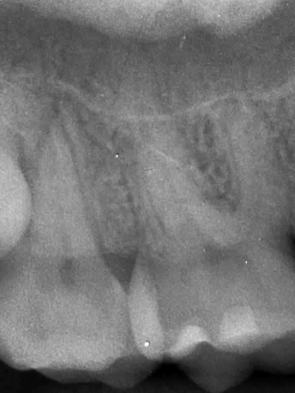
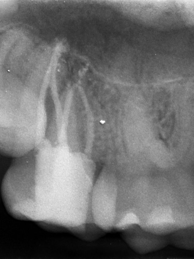
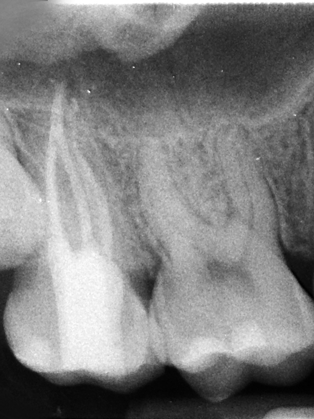
Conventional endodontic therapy of a maxillary second molar with atypical anatomy


Diagnostic and Treatment Considerations
- Position in the arch: 2nd or 3rd molars
- Crown and root morphology: Maxillary molar with atypical anatomy
The patient presented with a history of pain in the maxillary right quadrant. Clinical examination revealed that tooth #2 had a deep palatal groove and caries was present. Clinical testing supported a diagnosis of pulpal necrosis with symptomatic apical periodontitis. During endodontic treatment, a second palatal was located with the aid of a surgical operating microscope.
Although the patient was young and healthy, maxillary molars may offer significant anatomical variability. Due to their position in the arch, locating and negotiating all canals without supplementary magnification and illumination can be very challenging, especially if atypical anatomy is present.