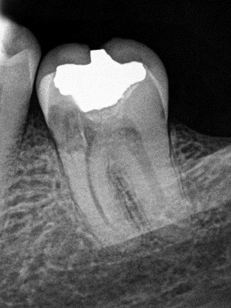
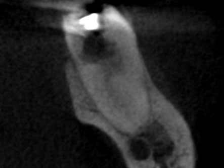
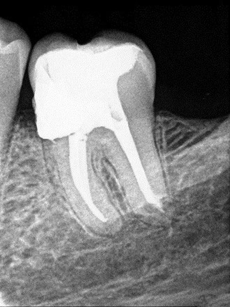
Diagnosis Patient presented with a non-vital pulp and symptomatic apical periodontitis. The preoperative radiograph displays an atypical root and canal configuration. The CBCT sagittal view shows that the mesiolingal and distal canals join and the mesiobuccal canal exits the root short of the root apex. Treatment After access opening, three canals were located and […]
Diagnosis Patient presented with symptomatic irreversible pulpitis and symptomatic apical periodontitis. The preoperative radiograph displays internal anatomy with a single canal that divides into three canals in the coronal third of the root. Treatment After access opening, three canals were located and instrumented. The postoperative radiograph confirms the filling of three canals with multiple […]
Diagnosis Patient presented with a non-vital pulp and chronic apical periodontitis. The pre-operative radiograph showed a radiolucent area surrounding the apical third of the root and advanced horizontal bone loss on the distal. The mesial root displayed a double curvature and there was a sharp dilaceration of the distal root with the canal exiting on […]
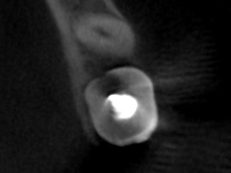
Diagnosis Patient presented with a non-vital pulp and chronic suppurative apical periodontitis. The preoperative radiographs showed aberrant anatomy that suggestive of a dens evaginatus. A gutta percha cone in the sinus tract traced to the distal aspect of the root and possible second canal. The CBCT axial view confirmed the presence of an additional root […]
Diagnosis Patient presented with previously initiated endodontic treatment. The preoperative radiograph disclosed that there was a separated instrument in the apical third of the mesial root. No other clinical signs or symptoms were present. Treatment Three canals were located and it was confirmed that a separated instrument was present in the mesiolingual canal. It was […]
Diagnosis Patient presented with a non-vital pulp and chronic suppurative apical periodontitis. The pre-operative radiograph showed a radiolucent area surrounding the apical third of the root. Treatment Three canals were located and instrumented. The distal root was not negotiable to the radiographic apex with hand or rotary files; so, the Sonendo GentleWave® procedure was […]
Diagnosis Patient presented with a non-vital pulp and symptomatic apical periodontitis. The preoperative radiograph displayed a widened periodontal ligament space associated with the mesial root apex. Treatment Four canals were located and instrumented followed by the Sonendo GentleWave® procedure to augment cleaning and disinfection. The postoperative radiograph confirms the filling of the four canals plus […]
Diagnosis Patient presented with a non-vital pulp and symptomatic apical periodontitis. The preoperative radiograph did not display any atypical findings. Treatment Three canals were located and instrumented followed by the Sonendo GentleWave® procedure to augment cleaning and disinfection. The post- operative radiograph displays an anastomosis between the mesial canals in the apical third of […]
Diagnosis Patient’s general dentist initiated endodontic treatment; but, was unable to locate any canals. Clinical testing confirmed the presence of a non-vital pulp and symptomatic apical periodontitis. The CBCT axial view displays three separate roots present in the apical third. Treatment Three canals were located and instrumented followed by the Sonendo GentleWave® procedure to […]
Diagnosis Patient presented with a non-vital pulp and asymptomatic apical periodontitis. The CBCT axial view confirms the presence of a “C” shaped root anatomy with three separate canals connected by ribbons of pulpal tissue. Treatment Three main canals were located and instrumented. This was followed by the Sonendo GentleWave® procedure to augment cleaning and […]
Diagnostic and Treatment Considerations Position in the arch: 2nd or 3rd molars Crown and root morphology: Maxillary molar with atypical anatomy The patient presented with a history of pain in the maxillary right quadrant. Clinical examination revealed that tooth #2 had a deep palatal groove and caries was present. Clinical testing supported a diagnosis […]
Diagnostic and Treatment Considerations Radiographic difficulties: Moderate difficulty interpreting radiographs Crown morphology: Significant deviation from normal tooth/root form (dens invaginatus) Patient presented with pain, tenderness to percussion, and intraoral swelling associated with tooth #7. Clinical testing supported a diagnosis of pulpal necrosis with symptomatic apical periodontitis. The radiographic examination revealed the presence of dens […]
Patient Considerations Medical History: Complex medical history/serious illness/disability Diagnostic and Treatment Considerations Position in the arch: 2nd or third molar; extreme inclination (> 30 degrees) Tooth isolation: Extensive pretreatment modification (restoration of distal caries) An elderly patient presented with a symptomatic mandibular second molar. Their medical history indicated that they were receiving a […]
Diagnostic and Treatment Considerations Position in the arch: Mandibular second molar Tooth isolation: Simple pretreatment modification required for rubber dam isolation Canal and root morphology: Extreme curvature (> 30°) or S-shaped curve Preoperative evaluation revealed extensive caries and a coronal fracture. Clinical testing supported a diagnosis of symptomatic irreversible pulpitis with symptomatic apical periodontitis. […]