

Diagnostic and Treatment Considerations
- Radiographic difficulties: Moderate difficulty interpreting radiographs
- Crown morphology: Significant deviation from normal tooth/root form (dens invaginatus)
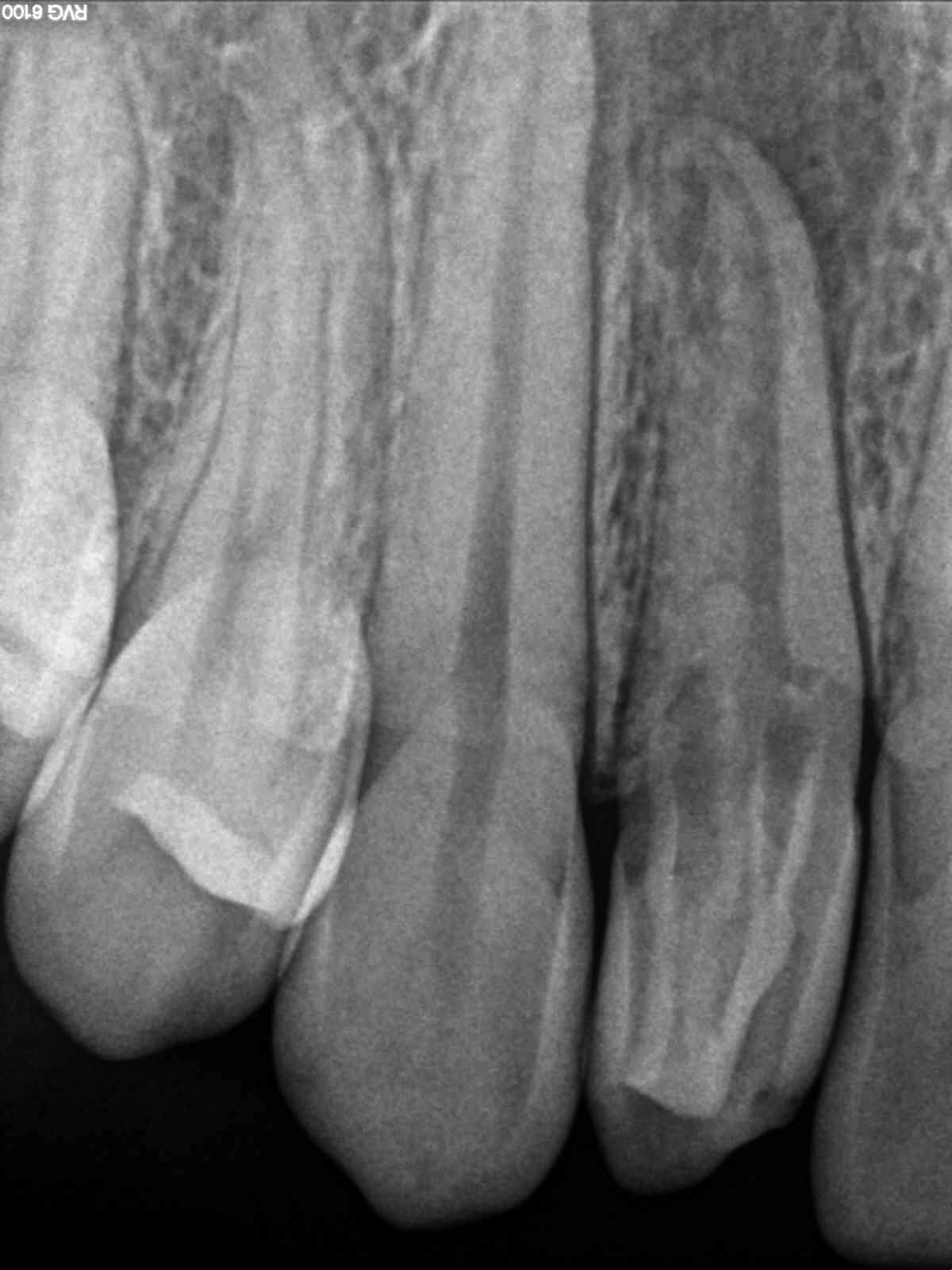
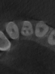
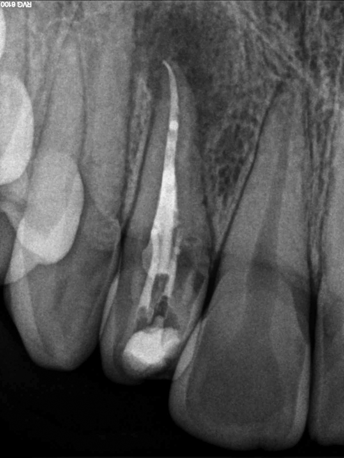
Patient presented with pain, tenderness to percussion, and intraoral swelling associated with tooth #7. Clinical testing supported a diagnosis of pulpal necrosis with symptomatic apical periodontitis. The radiographic examination revealed the presence of dens invaginatus (dens in dente) with two deep infoldings of enamel and dentin mesial and distal to a main canal. This finding was confirmed and better visualized for treatment planning with Cone Beam Computerized Tomography (CBCT).
While there were no patient considerations that exceeded a moderate level of difficulty, the atypical crown and root morphology associated with dens invaginatus makes these cases highly difficult to treat. Accurate pretreatment visualization and an understanding of the aberrant morphology are key factors in obtaining clinical success in cases such as this.