Maxillary lateral incisor with a second root


Diagnosis
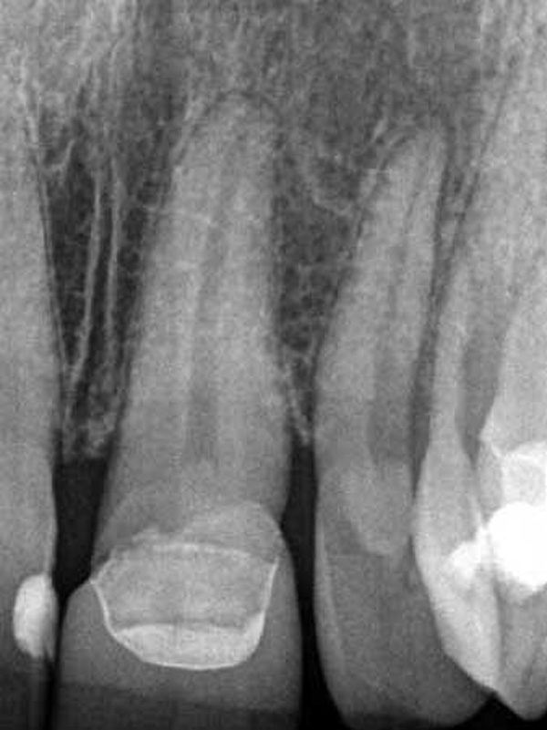
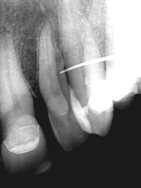
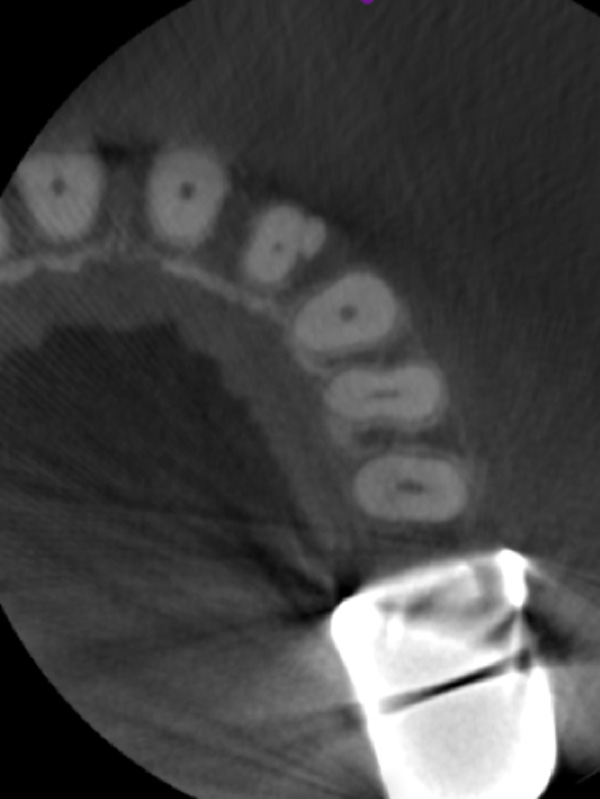
Patient presented with a non-vital pulp and chronic suppurative apical periodontitis. The preoperative radiographs showed aberrant anatomy that suggestive of a dens evaginatus. A gutta percha cone in the sinus tract traced to the distal aspect of the root and possible second canal. The CBCT axial view confirmed the presence of an additional root in the apical third.
Treatment
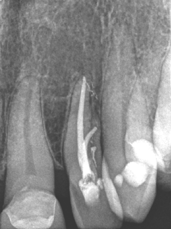
Two canals were located and instrumented. The postoperative radiograph confirms the filling of two roots and multiple portals of exit. At a three-month follow-up, the sinus tract had completely healed and the tooth was asymptomatic.