Studies around tooth morphology and the internal anatomy of root canal systems demonstrate great variations and complexities in the pulp space as it travels from the crown to the apex. Common findings include extra roots and canals, recesses, fins, isthmuses, accessory canals, and open apices. These are some of our best atypical anatomy cases, complete with diagnosis and approaches to treatment.
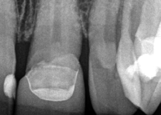
Case 01
Tooth #7: Maxillary lateral incisor with dens in dente.


Diagnosis
The fourteen-year-old patient presented with a non-vital pulp and symptomatic apical periodontitis. The preoperative radiograph displayed a periapical radiolucency associated with an incompletely formed root and dens in dente.
Treatment
Access to the middle and apical portion of the root was obtained by making an opening through the dens in dente. The canal space was cleaned and disinfected and calcium hydroxide was placed to augment disinfection and aid in root end closure (apexification). After medication changes and observation for six months, it was determined that apexification was taking place and the periapical radiolucency was decreasing in size. The canal space was filled by placing a collagen barrier at the apex and then filling the canal space with a bioceramic root repair material and gutta-percha.
Case 02
Tooth #10: Maxillary lateral incisor with a second root.


Diagnosis
Patient presented with a non-vital pulp and chronic suppurative apical periodontitis. The preoperative radiographs showed aberrant anatomy that suggestive of a dens evaginatus. A gutta-percha cone in the sinus tract traced to the distal aspect of the root and possible second canal. The CBCT axial view confirmed the presence of an additional root in the apical third.
Treatment
After access opening, three canals were located and instrumented. The postoperative radiograph confirms that the mesiobuccal and distal canals join and that there is an anastomosis between the mesial canals.
Case 03
Tooth #32: Mandibular molar with dilacerated root and canal exiting short of apex.


Diagnosis
Patient presented with a non-vital pulp and symptomatic apical periodontitis. The preoperative radiograph displays an atypical root and canal configuration. The CBCT sagittal view shows that the mesiolingal and distal canals join and the mesiobuccal canal exits the root short of the root apex.
Treatment
After access opening, three canals were located and instrumented. The postoperative radiograph confirms that the mesiobuccal and distal canals join and that there is an anastomosis between the mesial canals.