When a patient is experiencing pain and needs to be seen urgently, we make every attempt to add them to our schedule the same day. In fact, we see 25 emergent cases per day. Let us show you a few of our best:
Case 01
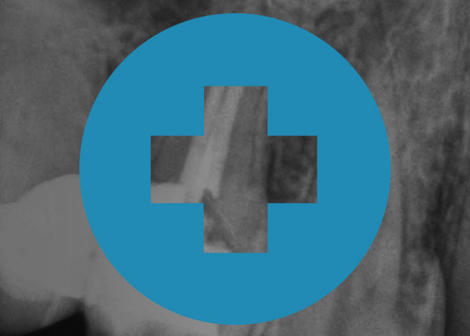
Tooth #4, Endodontic retreatment of a maxillary second bicuspid with overextended filling material
- Emergency condition: Severe pain
- Endodontic treatment history: Previous nonsurgical endodontic treatment with complications
Patient presented with severe pain in the maxillary right quadrant. Radiographic examination revealed previous nonsurgical endodontic treatment with the canal filling material extended beyond the apex and perforating the Schneiderian membrane.
Patient considerations such as severe pain requiring emergency care can create a high level of treatment difficulty. In addition, the history of previous endodontic therapy complicated by the overextension of canal filling material make the management of this case extremely challenging.

Case 02
Tooth #7, Maxillary lateral incisor with dens invaginatus.
- Radiographic difficulties: Moderate difficulty interpreting radiographs
- Crown morphology: Significant deviation from normal tooth/root form (dens invaginatus)

Patient presented with pain, tenderness to percussion, and intraoral swelling associated with tooth #7. Clinical testing supported a diagnosis of pulpal necrosis with symptomatic apical periodontitis. The radiographic examination revealed the presence of dens invaginatus (dens in dente) with two deep infoldings of enamel and dentin mesial and distal to a main canal. This finding was confirmed and better visualized for treatment planning with Cone Beam Computerized Tomography (CBCT).
While there were no patient considerations that exceeded a moderate level of difficulty, the atypical crown and root morphology associated with dens invaginatus makes these cases highly difficult to treat. Accurate pretreatment visualization and an understanding of the aberrant morphology are key factors in obtaining clinical success in cases such as this.
Case 03
Tooth #2, Conventional endodontic therapy of a maxillary second molar with atypical anatomy.
- Position in the arch: 2nd or 3rd molars
- Crown and root morphology: Maxillary molar with atypical anatomy

The patient presented with a history of pain in the maxillary right quadrant. Clinical examination revealed that tooth #2 had a deep palatal groove and caries was present. Clinical testing supported a diagnosis of pulpal necrosis with symptomatic apical periodontitis. During endodontic treatment, a second palatal was located with the aid of a surgical operating microscope.
Although the patient was young and healthy, maxillary molars may offer significant anatomical variability. Due to their position in the arch, locating and negotiating all canals without supplementary magnification and illumination can be very challenging, especially if atypical anatomy is present.
Scheduling
If you have a patient that is experiencing pain and needs to be seen urgently, please have them contact our office early in the day. Our extensive scheduling availability means they will receive the prompt and specialized attention they serve. Four locations, one great experience.