Treating anatomical aberrations requires thorough knowledge of expected anatomy and recognition of atypical variations.
There have been many studies regarding tooth morphology and the internal anatomy of root canal systems. They have all demonstrated great variations and complexities in the pulp space as it travels from the crown to the apex. Common findings include extra roots and canals, recesses, fins, isthmuses, accessory canals, and open apices. These anatomical aberrations may provide spaces for biofilms to reside and may contribute to persistent symptoms after endodontic therapy or possible treatment failure.
Some common findings include:

Better Diagnoses with CBCT
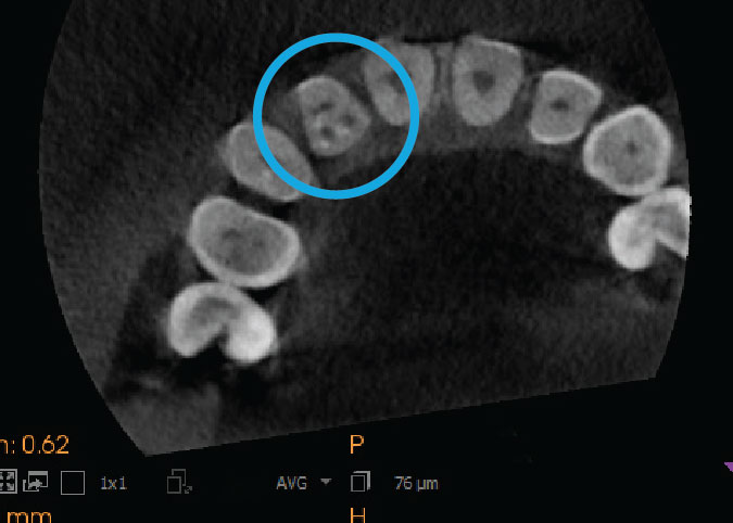
Radiographs are frequently used to help interpret canal variations; however, even with multiple angles, anatomical variations may be difficult to detect with 2D evaluations. The use of cone beam computed tomography (CBCT) has become a viable adjunct for clinicians to detect and visualize the canal system prior to treatment. Molar and premolar teeth usually present the highest incidence of variation. However, as exhibited in the CBCT below, anterior teeth may also display atypical morphology. Thorough knowledge of expected anatomy and variations from the norm are essential when undertaking endodontic therapy to improve success.

Let us show you our best work.
Visit Portfolio #04 to view and share some of our atypical tooth anatomy cases. All portfolios are available for download here.